


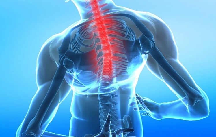
Spinal osteosarcoma is a complex of dystrophic and degenerative changes in the intervertebral discs and adjacent surfaces of the vertebral bodies associated with tissue destruction and disruption of their structure. Depending on the extent of the lesion, cervical, thoracic and lumbar osteonecrosis can be distinguished.
Symptom
The main signs by which one can assume the presence of osteonecrosis of the cervical spine is a local change in the configuration of one of the segments of the spine (development of scoliosis, scoliosis, scoliosis, scoliosis, and scoliosis). spine or scoliosis) - a clear curvature of the spine. in the vertical or horizontal plane. The second most common symptom is the pain syndrome, which can be localized not only to the vertebrae but also to other regions of the body by the corresponding nerve roots. Another complaint of these patients is discomfort and a feeling of neck fatigue.
With cervical osteochondrosis, as a rule, the pain manifests itself in the neck region and can reach the shoulder and shoulder blades, it can be confused with the pain of myocardial infarction, since it has symptoms. similar evidence. In addition, cervical osteonecrosis may be accompanied by frequent headaches and dizziness. When the arteries supplying the brain are compressed (squeezed), there may be signs of brain malfunction (neurological symptoms): fainting, nausea, ringing in the ears, mood swings, anxietysedimentation and other signs.
According to the severity of the pain, they are divided into 3 levels:
- Pain occurs only with pronounced movements in the spine;
- The pain is relieved by a certain position of the spine;
- The pain is permanent.
Forms
Depending on the syndrome caused in osteonecrosis, there are:
- Compression syndrome - occurs with compression (myopathy - compression of nerve roots, myelopathy - compression of muscles, nerve vessels - compression of blood vessels and nerves);
- Reflexes (muscle tonic, neuromuscular, neuromodulatory);
- Myoadaptive syndrome (overactivity of healthy muscles as they take over the functions of the affected muscles).
Reason
The mechanism of development of the disease is damage to the disc from various causes and its displacement along with the gradual loss of functions of the spine (mild pressure relief). Immediate causes of disc damage can be age-related degenerative changes related to impaired blood supply to the disc, mechanical damage from trauma and stress on the spine -overweight, for example.
An important role in the development of osteonecrosis is due to a sedentary lifestyle, in which a violation of the blood supply and function of the intervertebral joints develops. The mechanism of development of the disease is as follows: if the annulus connecting the vertebral bodies is damaged, the disc is pushed posteriorly - into the lumen of the spinal canal, or laterally - with the formation of medial and lateral discs. herniation. Discs can be pushed into the vertebral body with the formation of Schmorl hernias - microscopic breaks of the cartilage tissue of the disc into the spongy tissue of the vertebrae. In the case of a posteriorly displaced disc, compression of the spinal cord and the roots extending from it is possible, with the development of the typical pain syndrome.
Diagnose
Diagnosis of osteonecrosis of the spine is made on the basis of complaints, pathological data, clinical examination and instrumental examination methods. Diagnostic measures are to find the cause that led to the development of neurological symptoms.
From the medical history, it is possible to find out the presence of injuries, the nature of work - constant physical overload (lifting weights), wrong posture, peculiarities of work and the position of the upper spinetable and while walking, the presence of infection.
General clinical studies (clinical blood tests, general urinalysis), blood biochemistry tests have no independent value. They are prescribed to assess existing conditions, diagnose underlying disease, and emergent complications.
Diagnosis is based on the clinical picture of the disease and is made by sequential exclusion of diseases that are similar in clinical signs. Among the instrumental diagnostic methods, the most common and available is the x-ray examination (spondoscopy is a non-contrast study). It reflects the narrowing of the intervertebral disc space and allows you to identify osteoclasts (bone growths) on the vertebral bodies, but provides only indirect information on the extent of damage to the vertebral bodies. disc.
Accurate diagnosis can be made by CT and MRI imaging (computed and magnetic resonance imaging), even in the early stages of the disease. CT allows you to identify minimal abnormalities in bone and cartilage tissue, MRI - to perform detailed soft tissue structures and determine the location of a herniated disc.
A duplex ultrasound scan of the cerebral arteries is done if a violation of the blood supply to the brain is suspected.
Differential diagnosis is carried out with diseases with similar clinical manifestations: progressive pathology with pain radiating to the shoulder and squamous area (liver disease, gallbladder disease, pancreatitis - pancreatitis); cervical lymphadenitis - an increase in the lymph nodes in the cervix, rheumatoid arthritis; cancer (tumors of vertebrae, roots, spinal cord and membranes), tumors of the pharynx and pharynx, Pancost cancer (brachial plexus compression in upper lobe lung cancer), neck metastasesuterus; tuberculous spondylitis - an inflammatory spondylitis caused by mycobacterium tuberculosis; arachnoid cyst; pseudocyst of the dura mater; spinal deformities; fibromyalgia is a disease that causes pain in the muscles, ligaments, and tendons, thoracic outlet compression syndrome - a disorder caused by excessive pressure on the bundle of nerve vessels that pass between the anterior and medial muscles, above the ribs. first and subclavian, syndromic and scapular muscles - a chronic pathological condition caused by the formation of local spasm or sealing muscles, manifested by painful spots.
The main laboratory tests used:
- Clinical blood test;
- Blood chemistry.
The main instrumental studies used:
- X-ray of the spine (spondylography);
- Magnetic resonance imaging (MRI);
- Computed tomography (CT);
- Double-sided ultrasound scan of the cerebral arteries (if a violation of the blood supply to the brain is suspected).
Additional instrumental studies used:
- Densitometry - measuring bone density (as indicated).
Treatment
Treatment of osteonecrosis of the spine completely depends on the stage and degree of development of the osteonecrosis. In the early stages, it is possible to use preventive measures, physical therapy, simulated movements, and gymnastics. With severe pain syndrome, the patient needs to rest the body. Anti-inflammatory and antispasmodic drugs are prescribed. It is possible to block the disc with anesthetic to open the pathological ring, when the pain will cause muscle spasm, and at the same time the disc is compressed more strongly, thereby increasing the pain itself.
Warming ointments are applied topically to the skin over the vertebral column to improve local blood supply and reduce tissue edema. These patients are shown wearing corsets. In patients with an early stage of the development of osteonecrosis, chondroprotectors are effective - drugs that improve the restoration of cartilage tissue, as well as drugs that improve local blood supply, venotonics, vitaminsgroup B. In the case of pain syndrome does not stop. Medically long time and there is spinal root compression clinic with herniated disc, surgical removal of damaged disc is indicated. In cases where the disc compresses the entire spinal cord, early surgery is indicated.
You shouldn't wait until a person begins to urinate or defecate spontaneously - in this case, the spinal cord damage may already be irreversible. As physiotherapeutic procedures, magnetic therapy, ultrasound, massage, manual therapy, acupuncture and physiotherapeutic exercises are prescribed.
Complications
There may be vegetative-vascular dystonia and cardiac disruption, cerebrovascular accident, hypotension and hypertension (decreased and increased blood pressure), vestibular disorders (impaired coordination of movements). ), vertebral artery syndrome (a disease caused by narrowing of the vertebral arteries), periarthritis (a disease that impairs mobility) of the shoulder joint.
Preventive
To prevent osteonecrosis, it is necessary to address the factors that cause the disease, which are: avoiding spinal injury, stress on the spine (lifting weights), preventing overweight. For people who already have the early stages of osteonecrosis, a corset should be worn at home and during exertion. In order for the spine to rest during sleep, you should sleep on an orthopedic mattress and pillow.
What questions should you ask your doctor?
Are there exercises to help relieve symptoms?
What drugs will help to cope with cervical osteochondrosis?
What happens if you don't get treatment in time?
Patient advice
Exercise, weight loss when overweight, cool or warm compresses help reduce the symptoms of thoracic spondylosis. It's important to eat right, monitor your spine, treat chronic conditions, and avoid injury.